As of 2024, there are no successful medications that remove pterygiums. Surgery remains the gold standard for pterygium removal. During pterygium surgery, the small fleshy, triangular growth of tissue on the surface of the eye is removed and replaced with an autograft, a healthy piece of conjunctival from the patient’s own eye transplanted into the former area of the prior pterygium to act as a barrier. Here’s an overview of pterygium surgery in more detail:
- Preoperative evaluation: Before undergoing pterygium surgery, you will have a comprehensive eye examination to assess the extent of the pterygium, evaluate your overall eye health, and determine your suitability for surgery.
- Anesthesia: Pterygium surgery is usually performed under local anesthesia, which numbs the eye and surrounding tissues. In some cases, the surgeon may recommend mild sedation or general anesthesia depending on the patient’s preference and the complexity of the procedure.
- Pterygium removal: The surgeon carefully removes the pterygium tissue from the surface of the eye. This involves making an incision around the pterygium and dissecting and removing the abnormal tissue. The underlying healthy conjunctiva may be repositioned and sutured into place to cover the affected area.
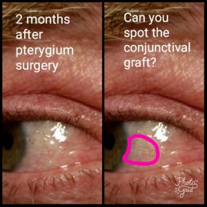
- Graft placement: In some cases, to prevent the pterygium from regrowing, the surgeon may use a graft to cover the area where the pterygium was removed. The graft is usually taken from the patient’s own conjunctiva, or in some cases, it may be a synthetic or amniotic membrane graft. The graft is secured in place with sutures.
- Postoperative care: After the surgery, you will be prescribed eye drops or ointments to help with healing and prevent infection. It is important to follow the postoperative care instructions provided by your surgeon, including the use of prescribed medications and any activity restrictions. Your surgeon will schedule follow-up visits to monitor the healing process and remove sutures if necessary.
- Recovery and healing: The recovery time after pterygium surgery can vary from a few weeks to several months. Initially, you may experience some discomfort, redness, tearing, and blurry vision. It is normal for the eye to appear red and swollen during the healing process. It’s important to avoid rubbing or scratching the eye, and you may be advised to wear a protective eye patch or shield during the initial healing period.
It’s worth noting that while pterygium surgery can effectively remove the growth and alleviate symptoms, there is a possibility of recurrence. To minimize the chances of recurrence, your surgeon may recommend postoperative measures such as the use of eye drops or medications and the regular use of protective UV eyewear.
The reported rates of pterygium recurrence after surgery vary in the literature, ranging from around 4-10%. The use of autografts, a healthy conjunctival transplant from the patient’s own eye placed at the prior site of the old pterygium, can prevent recurrence. In comparison, older techniques like bare sclera excision (without a graft) have much higher recurrence rates, sometimes exceeding 30% to 50%. The conjunctival autograft is generally considered the most effective and safest option for reducing recurrence.
As with any surgical procedure, pterygium surgery carries potential risks and complications, including infection, scarring, bleeding, dry eye, and changes in vision. It is important to consult with an experienced ophthalmologist or eye surgeon who can evaluate your specific condition, discuss the potential risks and benefits, and determine the most appropriate treatment approach for you.
This article was originally written on 5/29/2023 by Angelique Pillar, MD and updated 9/15/2024.